This case was reported in the Saint Bartholomew’s Hospital Reports – the in-house journal published by the London hospital of the same name – in 1879. The author of this article, William Steavenson, was a 29-year-old house physician at Barts (as those familiar with the hospital call it). Steavenson’s interests included chronic asthma – from which he had suffered since childhood – and the uses of electricity in medicine, about which he wrote a textbook which was published on both sides of the Atlantic. He died at the tragically early age of 41 – killed by influenza, a vulnerable person during another terrible pandemic.

J.D., aged 21, a gentleman’s groom, was admitted into St. Bartholomew’s Hospital on April 23, 1879, recovering from right hemiplegia.
Hemiplegia is a form of paralysis affecting one side of the body. It is most often caused by injury to the brain or (sometimes) spine.
About sixteen months before, at his master’s residence in the country, when larking in the servants’ hall, he attempted to kiss the kitchen-maid.
An act that would not be described as ‘larking’ by many employers today. The question of whether the maid had the slightest interest in being kissed by the groom is not addressed.
This young person had been engaged in knitting, and had one of the knitting-needles placed behind her left ear.
Can you guess what happened next?
As the face of the groom approached that of the young woman, the knitting-needle entered his left orbit, passing between the bone and the eyeball, to the depth, it is said, of four inches, no doubt entering the brain.
Or around 10 cm. Quite a long way into the brain, in all likelihood.
It must be supposed that the ardour of the young man was extreme, and that most likely the needle obtained a point d’appui in the lady’s back hair…
A point d’appui (literally, ‘fulcrum’) is a military term meaning a base or strong point.
…but such was the confusion following the occurrence that the history on these points is not very clear; but this much is certain, that J. D. retired from the encounter with the knitting-needle protruding from his orbit.
A vignette of extreme horror or high slapstick, depending on your point of view.
He says he removed it with his own hand.
Not, in general, a good idea. If in doubt, leave a foreign object in place and let a doctor take it out for you.
It was followed by some bleeding, and he then fainted.
Understandably.
We therefore have no accurate knowledge of the exact direction of the implement, but, from the symptoms which followed, it is very probable that it passed through the sphenoidal fissure, and must ultimately have injured the brain somewhere in the neighbourhood of the third left frontal convolution, or what is called Broca’s convolution.
The sphenoidal or superior orbital fissure is a gap between two bones at the back of the eye socket. More interesting, however, is the reference to ‘Broca’s convolution’ (known today as Broca’s area), a region of the left frontal lobe of the brain.
In the 1860s a physician in France, Pierre Paul Broca, encountered several patients with aphasia (inability to speak) who all turned out to have lesions in the same region of the brain. This was the first convincing evidence for the localisation of brain function – the idea that particular areas of the brain perform specialised tasks. Experimental work over the following decades established that other parts of the brain were associated with other functions such as locomotion and sensory abilities.
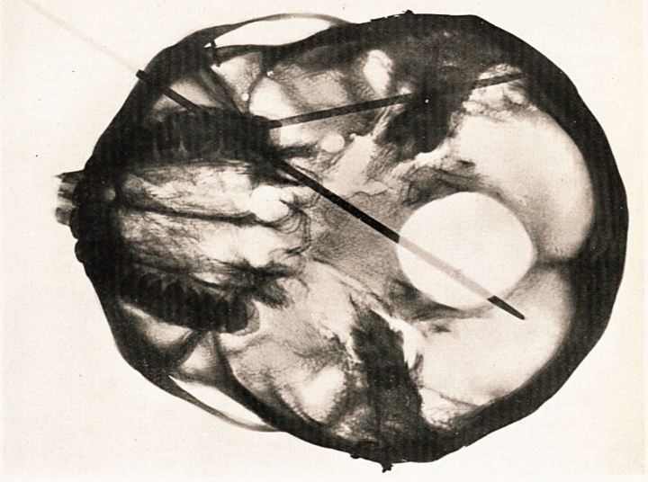
In the hospital post-mortem room, Dr Steavenson performed experiments on several cadavers, passing knitting needles through their eye sockets to find out which parts of the brain might be injured, and found that it was quite feasible for a needle to reach Broca’s area. Half a century later the Edinburgh surgeon David Greig performed a similar experiment (using a skull rather than a complete cadaver) and X-rayed the results, demonstrating the range of angles at which a knitting needle might enter the brain via this route:
As for Dr Steavenson’s patient:
The injury at the time caused the patient intense pain, but did not injure his sight. Up to the fifth day after the accident he could talk and use his right arm and leg. Then he lost all power over his right upper extremity, side, and leg. The lower part of the right side of his face was paralysed, and he could not whistle. The right side of the tongue was also paralysed. The paralysis came on at night. At first the paralysed parts felt cold and dead, but he did not lose sensation. He lost the power of speech for two months.
The symptom observed by Broca in his patients, known clinically as aphasia.
On his admission to the Salop Infirmary at Shrewsbury a short time after the accident, he could only say the words “Yes, yes,” which he answered to every question.
Broca’s first patient could only utter the syllable ‘tan’, and would respond to any question with the phrase ‘tan, tan’.
The almost total paralysis remained as long as the aphasia, viz., for two months. He never lost power over his sphincters. He is now gradually recovering power over all the paralysed parts. Occasionally he has severe aching in the back of his head and on the left side of his neck, which lasts for some hours. His memory is good. He has had three epileptiform fits since the accident, one in December 1878, one in February of this year, and one in April a few days before admission.
The patient was, it seems, somewhat improved by the time he was transferred to Bart’s.
On admission, he had the appearance of being a strong, robust man in perfect health, with a florid complexion. He complained of inability to use his right hand or fingers, with weakness and only partial power over his right side, arm, and lower extremity…He remained under observation about five weeks, with galvanism applied to his arm and hand…
Galvanism is the medical use of electricity, often used at this date to treat paralysis. Since it had long been known that muscle fibres contracted when a current was passed through them, it seemed logical to apply electricity in this way. Dr Steavenson was an acknowledged authority on the subject, appointed the first head of the Barts Electrical Department on its foundation in 1882.
…the hand placed in the intervals upon a splint to try and bring the fingers straight, but very little improvement was observed in his condition from the time when he entered the Hospital. The contraction and rigidity of the flexors of the forearm were of too old standing to expect much improvement. He had no fits while under treatment
The human brain is a remarkable thing, but its ability to heal after physical injury is limited. There was nothing the doctors could do for this patient beyond careful rehabilitation, but he still seems to have recovered most of the function he lost in the immediate aftermath of the accident. The fact that he even survived four inches of knitting needle inside his skull is pretty extraordinary.