The Northern Journal of Medicine was a short-lived periodical which appeared for only two years before being acquired by a more successful competitor. But it had some illustrious contributors: published in Edinburgh, it was able to include papers by some of the most eminent medical academics in Europe. The very first edition, which appeared in May 1844, included this article by James Duncan, a 33-year-old surgeon at the Edinburgh Royal Infirmary:

—— Calder, aged 22, Edinburgh, March 9, 1844, had lost his two superior anterior incisors when a boy by an injury received when playing at ball.
The boy had knocked out his two upper front teeth. Today a dentist would call these the maxillary central incisors.
Being a dentist’s workman, he had, to conceal the loss, made at his spare hours artificial substitutes which he had worn for the last three months.
This almost seems too convenient. One wonders whether his missing front teeth influenced his choice of career.
These were intended to be removed at night, and in consequence of this, as well as to prevent them from being seen, he had made the gold plates, by means of which they were attached to the adjoining teeth, as little projecting as possible, compatible with the retaining them in position. The consequence was, that their grasp of the adjacent teeth was but slight, and they could be displaced with ease.
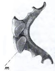
The article includes a small sketch of the item in question, which looks much like a modern partial denture – though today’s models are unlikely to use gold plates.
His master had repeatedly warned him of the danger attending the wearing them during sleep, and recommended him always to remove them. He had, however, neglected this precaution, and on the 28th February last, he had gone to rest with them as usual.
Can anybody guess what happened next?
In the morning the teeth were missing; and after a fruitless search for them amongst the bed-clothes, he became convinced that he must have swallowed them. He was further confirmed in this opinion by the difficulty in swallowing which he experienced, and by the sharp pain which he suffered when the attempt was made.
It does not require the diagnostic skills of Gregory House MD to work out what had happened.
He was naturally much alarmed by the accident, and in consequence applied to Mr Syme for assistance.
James Syme was a great Edinburgh surgeon also notable as the inventor of the waterproofing process used to create the mackintosh.
Mr S. passed a probang, and detected a foreign body in the oesophagus, considerably below the cricoid cartilage, and much beyond the reach of the ordinary forceps used for extracting foreign bodies from the gullet.
A probang is a long flexible rod with a sponge at the end, used to push foreign bodies from the oesophagus into the stomach. The cricoid cartilage is below the Adam’s apple, which gives you some idea of how far down this object was stuck.
Mr S. now recommended his removal to the hospital, and when there introduced a probang with threads passed through the bulb, the other ends being retained in the hand, trusting that if the bulb could be carried beyond the foreign body, it might be entangled by them, and thus removed. Nothing, however, was detected, and it was believed that it had found its way into the stomach,—an opinion which was rendered the more probable by the fact, that the difficult deglutition was by no means so great as previously.
He no longer had trouble swallowing, in other words.
So considerable indeed was the relief, that the young man requested permission to leave the hospital the same evening. It was thought unsafe to comply with this request, and he remained in hospital nine days, still suffering from the fixed pain already alluded to, and occasionally spitting small quantities of blood, but without complaining of much difficulty in deglutition.
Spitting blood is obviously a bad sign, and one which I imagine would prompt urgent investigations today. But the surgeons of 1844 had no X-rays, endoscopes or other means of internal imaging. They believed the bleeding was trivial, and interpreted the pain as a residual symptom of the awkward passage of the false teeth through the oesophagus. They thought it unnecessary or dangerous to try any further intervention, and the patient went home. Prematurely, as it turned out.
Next morning, the 9th of March, I received a hurried call from his mother, who had been much alarmed by an occurrence which had taken place shortly before I had been sent for. She stated that her son had risen from bed, and that in crossing the room towards the window, he had become suddenly faint and giddy, and had vomited a mouthful of blood. He was immediately removed to bed, and complained of a feeling of great weakness.
Dr Duncan hurried to the man’s bedside, and found him weak, pale and struggling to breathe.
From his description of what had taken place, I was led to believe that the foreign body had been dislodged from its situation, and that it was possibly within reach of the forceps, with which I had provided myself. I accordingly requested him to sit up by the side of his bed, to enable me to make the necessary examination. This he did with ease, and without much assistance, expressing great anxiety to have something done to relieve him.
Dr Duncan was proved correct in his assumption that the false teeth had finally been dislodged. But his attempt to remove them was tragically misguided.
The act of depressing the tongue, to enable me to introduce the forceps, produced vomiting, and a mouthful of dark fetid blood was discharged. This was immediately followed by another but much larger quantity of fluid of the same description, perhaps about eight or ten ounces, and the false teeth were heard to rattle against the vessel into which it was received.
Good news: the young man had finally managed to vomit up the denture. Bad news: he had lost more than half a pint of blood in doing so.
The patient was immediately aware of this, and his friends were overjoyed at what had taken place. Another mouthful of the same fluid was then ejected; an interval of a few seconds elapsed, and then a mouthful of bright arterial blood was discharged; a second, and a third followed, the lips became livid, the pulse at the wrist ceased, the patient gave one or two convulsive sobs, and expired.
A brutally unexpected twist in the narrative. It is clear from the doctor’s account that the man’s friends assumed that since the teeth had been ejected, he would soon be getting better. As Dr Duncan immediately recognised, the denture had perforated a large artery of some description; but until the autopsy he had no idea which.
The oesophagus, stomach, and duodenum were found distended with pretty bright arterial blood. The quantity could not be measured; but in the opinion of those present at the examination, Professor Henderson, Mr Shand, Mr Reid, and myself, there could not be less than eight or ten pounds.
This is a vast amount of blood – many pints, and easily enough to cause rapid death.
The pharynx and oesophagus were laid open by an incision posteriorly, carried as low as the cardiac orifice of the stomach. About 4½ inches from the rima glottidis there was an ulcerated perforation of the anterior part of the oesophagus, of about ¾ths of an inch in length and three lines in breadth, passing obliquely upwards from the right to the left side.
The rima glottidis is the narrowest part of the cavity of the larynx. The false teeth had caused a large opening in the gullet some distance below it.
By this opening the probe could be readily passed into the aorta; but the latter vessel was not laid open at the time, it being thought better to immerse it for a day or two in spirits before doing so. On laying open the aorta subsequently, a perforation of about the size of a large crowquill was found about half an inch below the origin of the left subclavian artery.
The denture had caused a small hole in the aorta – but that was all that was needed to kill him very quickly indeed. The aorta, the largest artery in the body, contains blood at extremely high pressure. Even a tiny hole in it can cause catastrophic haemorrhage.

The gold plate to which the teeth were attached was pretty large, adapted to the shape of the palate immediately behind the incisor canine and bicuspid teeth on either side, with projections corresponding to the spaces between these teeth. The two last of these projections on both sides were large and pointed, with almost a cutting edge.
The surgeon observes that
one could scarcely conceive a more dangerous weapon, or one more likely to be followed by fatal consequences, than that I have just described, when lodged in the situation in which this one must have remained from the time of the accident.
It’s not often that dentures are described as a ‘dangerous weapon’. But in this case it seems the comparison was justified.